
Cardiogenic pulmonary edema in dogs is a serious condition where fluid accumulates in the lungs due to left-sided congestive heart failure (CHF). This occurs when increased pressure in the pulmonary capillaries (from backup of blood caused by heart dysfunction) forces fluid out of the blood vessels into the lung tissue and air sacs (alveoli). It is distinct from non-cardiogenic pulmonary edema, which stems from other causes like trauma, seizures, electrocution, or inflammation without primary heart failure.
Common Causes
The most frequent underlying heart diseases leading to cardiogenic pulmonary edema in dogs include:
- Myxomatous mitral valve disease (also called degenerative or chronic valvular disease/endocardiosis): Especially common in small-breed, older dogs; the mitral valve leaks, causing blood to flow backward and increasing left atrial pressure.
- Dilated cardiomyopathy (DCM): More common in large-breed dogs; the heart muscle weakens and enlarges, impairing pumping efficiency.
- Other contributors: Hypertrophic cardiomyopathy (less common), congenital defects, or factors like high-sodium diets that can exacerbate fluid retention.
For cardiogenic edema to develop, there must be identifiable left-sided heart disease causing elevated hydrostatic pressure in the pulmonary capillaries (normally <12 mmHg).
Symptoms
Dogs often present as an emergency with:
- Rapid or labored breathing (tachypnea or dyspnea), sometimes with orthopnea (preferring to stand or sit with neck extended).
- Coughing (though cough is more commonly linked to primary lung issues than pure edema).
- Weakness, exercise intolerance, or collapse.
- Bluish gums or tongue (cyanosis) in severe cases.
- History of a heart murmur (common with mitral valve disease).
Many dogs have a prior diagnosis of heart disease, but edema can be the first obvious sign of decompensation.
Diagnosis
Veterinarians diagnose this through a combination of:
- Physical exam: Listening for heart murmurs, gallops, or arrhythmias; assessing respiratory rate and effort.
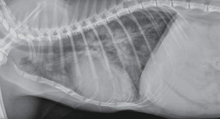
- Thoracic radiographs (X-rays): Hallmark findings in dogs include left-sided cardiomegaly (enlarged heart, especially left atrium), dilated pulmonary veins, and an interstitial-to-alveolar lung pattern starting in the perihilar (central/hilar) region and progressing to the caudodorsal (rear upper) lung fields. The pattern is often symmetric.
- Echocardiography (heart ultrasound): Confirms the underlying cardiac disease (e.g., valve regurgitation, chamber enlargement, poor contractility).
- Lung ultrasound: Can quickly detect “B-lines” (ultrasound artifacts indicating fluid) at the bedside for monitoring.
- Other tests: Bloodwork (e.g., NT-proBNP levels may be elevated in CHF), blood pressure, and ruling out non-cardiogenic causes.
Response to a trial of diuretics (e.g., reduced respiratory rate after furosemide) can support the diagnosis of cardiogenic origin.
Treatment
This is a medical emergency requiring immediate stabilization:
- Oxygen therapy: Via mask, cage, or nasal cannula to improve oxygenation.
- Diuretics: Furosemide (Lasix) is the cornerstone—typically 2–4 mg/kg IV/IM initially, repeated as needed (or as a constant-rate infusion). It reduces fluid overload by promoting urine output. Torsemide is sometimes used as an alternative.
- Rest and minimal stress: Handle gently to avoid worsening distress.
- Vasodilators: Such as nitroglycerin ointment (to reduce preload) or others like nitroprusside/hydralazine in severe cases (with blood pressure monitoring).
- Additional support: Pimobendan (to improve heart contractility), ACE inhibitors (e.g., enalapril or benazepril) for long-term management, and sometimes positive inotropes or mechanical ventilation in critical cases.
Once stable, address the underlying heart disease with lifelong medications, a low-sodium diet, and regular monitoring.
Prognosis
The immediate prognosis for resolving an acute episode is often good with prompt treatment, but long-term outlook depends on the severity of the underlying heart disease. Many dogs require ongoing therapy and can live comfortably for months to years with good management. Recurrence is possible, so owners should monitor resting respiratory rate (normal is typically <30 breaths/min at rest) and seek vet care at early signs. Severe or refractory cases carry a more guarded prognosis.
Important note: This is general information based on veterinary medical knowledge. Cardiogenic pulmonary edema is a life-threatening emergency—contact your veterinarian or an emergency clinic immediately if your dog shows signs of respiratory distress. Diagnosis and treatment must be tailored by a licensed veterinarian, often involving a cardiologist for complex cases. Early intervention improves outcomes significantly.